
Key Takeaways
- Identify persistent fever, spreading redness, worsening pain, foul discharge, or new hard lumps post-BBL as potential infection symptoms and consult a doctor immediately.
- Monitor temperature daily and record wound changes such as color, size, drainage, odor, and pain patterns to identify early symptom escalation.
- Know the difference between normal healing and infection. Normal healing will show steady improvement in soreness and skin coloration, whereas infection will present with worsening or new systemic symptoms such as chills and malaise.
- Anticipate physicians to employ physical examination, wound cultures, blood tests and imaging to diagnose infection and direct therapy.
- Know that treatment might need specific antibiotics, incision and drainage, or debridement. Post treatment wound care and observation are vital to healing.
- Minimize risk by selecting board-certified surgeons, guaranteeing sterile technique and postoperative care, and quickly reporting any alarming or fast-changing symptoms to a medical professional.
Bbl infection symptoms Typical symptoms are fever, spreading redness, warmth, pus and escalating pain at the incisions.
Additional symptoms include swollen lymph nodes, unexplained fluid drainage or lingering fatigue and chills.
Early medical review is important because infections can spread or involve implants and tissue viability.
The torso covers causes, timelines, when to seek care, and treatment.
Warning Signs
Warning Signs Early after BBL, inflammation and moderate pain peak in the first week. The body’s normal healing encompasses pain, which typically peaks around 48 to 72 hours and abates over the subsequent 2 to 3 weeks.
Still, some changes require immediate action as infections typically manifest within 5 to 10 days and necrosis signs can show up 1 to 2 weeks post-operation. Below these areas, we break down the key warning signs, what to watch for, and how to record observations for your care team.
1. Persistent Fever
Monitor for fevers greater than 38°C (100.4°F) for more than 24 hours post-BBL as a red flag. Follow temperature twice a day and record.
Persistent or climbing temperatures are more worrisome than an isolated spike. Chills, night sweats, or just generally feeling sick can accompany infection and can be an indication of spread throughout the system beyond the local wound.
Warning signs of deeper soft tissue infection or abscess that require imaging or drainage include persistent fever. Record exact temperatures, time of day, and any medication taken. This record helps clinicians decide whether antibiotics, imaging, or urgent evaluation is needed.
2. Spreading Redness
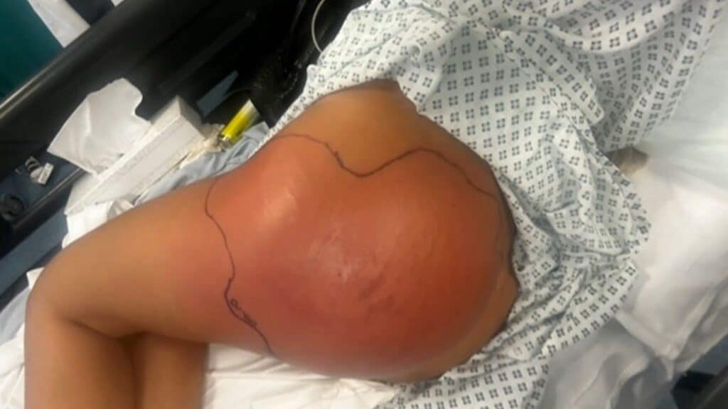
Look out for redness that extends outside incision lines into surrounding gluteal or thigh skin. Expanding erythema usually indicates cellulitis.
Warmth, swelling, and tenderness in those areas often go hand in hand with inflammation and indicate infection instead of normal bruising. Check both buttocks and adjacent thighs for symmetry.
Asymmetric or unilateral spread often helps localize an infectious process. Mark and photograph red zones every day, recording dimensions and any streaking toward lymphatic channels.
Note color changes. If red turns purple or dusky, and any areas spread quickly, contact your surgeon immediately.
3. Foul Discharge
Watch for pus, cloudy fluid, or yellow-green drainage from wounds or occult incision sites. Any new or worsening drainage, or drainage associated with skin breakdown, is concerning.
A stinky and/or “funky” smell is usually a sign of bacteria or an abscess pocket. Observe drainage color, consistency, and odor, and retain dressings for provider evaluation if needed.
Increased drainage with delayed wound healing should prompt culture and targeted antibiotics when indicated.
4. Escalating Pain
Pain that aims to relieve itself during convalescence deserves an examination. Normal post-op pain that peaks at 48 to 72 hours and then dissipates, any departure from that pattern is a red flag.
Pain radiating to the thigh, pelvis, or abdomen, or sharp and throbbing pain can sometimes be a sign of deep tissue involvement such as fat necrosis or abscess. If pain restricts walking or sleep, seek care.
Bring a pain log with ratings and pain provokers to help your doctor diagnose.
5. Hard Lumps
Be aware of hard, immobile lumps beneath the skin in the buttocks, lateral hip, or proximal thigh as these can be fat necrosis, seromas, or abscesses. New lumps that appear one to two weeks post-op warrant attention.
Watch lumps for growing larger, becoming painful, or developing changes in the overlying skin. Painful, enlarging nodules frequently require ultrasound or aspiration.
Record site, millimeter size, and any associated symptoms in an easy-to-reference table for physicians.
Critical Distinctions
Why it matters to differentiate normal recovery from active infection after BBL. Here are definitive signs to distinguish what is normal and what needs immediate medical attention.
Normal Healing
Anticipate moderate swelling, bruising, and soreness in the buttocks and adjoining regions for a few days to weeks after surgery. These signs typically reach their apex within the first 48 to 72 hours and then subside.
Pain should become more manageable by the day with standard pain medication. Some hardness or lumps are expected as transferred fat settles and re-integrates. Lumps tend to soften over the course of weeks.
Oil cysts can arise later in the healing process, usually between two and four weeks, and may present as smooth, movable nodules. Incision sites should show steady improvement. Skin color returns toward normal and any superficial scabs heal without increasing redness.
Maintain your regular post-op care like wearing compression garments, not sitting on your buttocks, and keeping your wounds clean and dry. Routine follow-ups allow the surgeon to observe fat take, seroma formation, and any early signs that the anticipated course is not being followed.
Active Infection
Persistent or increasing swelling, spreading redness or increasing pain—particularly if one side is worse—indicates away from normal healing and toward infection or other complications. Pain on one side or on a focal spot can indicate a hematoma, seroma, or if severe and rare, fat embolism.
Treat unilateral worsening seriously. Check for unusual wound discharge, offensive odor, or wounds that don’t heal. These are all strong signs of infection. Uneven swelling, early redness, or fever within 24 to 48 hours are red flags.
Seromas are the most frequent snag, comprising about 1 to 2 percent of complications and can get infected. Cellulitis is rare, occurring in less than 1 percent of cases, but can happen after liposuction skin breaks.
Extreme complications such as soft tissue abscesses, ulceration, or tissue necrosis can occur. Any skin changes, such as new numbness, persistent induration, red bumps at injection sites, or lumps that do not resolve between weeks three and six, are red flags and can be harbingers of permanent nerve damage or abnormal scarring.
Infections with non-tuberculous mycobacteria like M. Abscessus, which are notoriously difficult to eradicate, can necessitate months of focused antibiotic treatment. Act rapidly if systemic symptoms including fever, chills, or malaise accompany local signs.
Early treatment decreases the likelihood of permanent damage. Call your surgeon or urgent care if any of the above signs emerge instead of holding out for them to improve on their own.
Hidden Risks
BBL has hidden risks. Bad hygiene, lack of proper sterilization, or poor surgical technique increase the risk of infection and other complications. If instruments, implants, or the operating field are not clean, bacteria can infiltrate small incisions or injection sites. Clinics that skimp on sterilization or crews that hurry prep endanger patients. Antibiotic treatment may be used to prevent or treat infection, but it does not substitute for safe technique or proper wound care.
Fat embolism is among the most dangerous hidden risks. This occurs when fat is forced into veins and is carried to the lungs or brain, resulting in sudden breathlessness, oxygen deprivation, or worse. Symptoms may arise hours to days post-surgery. Expert surgeons mitigate this risk by not injecting into deep muscle layers and reserving fat grafts to the subcutaneous plane.
Fat necrosis, which is fat that dies and creates hard lumps or oil cysts, can appear later as firm nodules or pain and may need to be drained or operated on. Certain issues progress insidiously or without obvious early symptoms. Deep tissue infections can smolder beneath the skin without redness or fever initially, particularly in immunocompromised individuals.
Immune suppression from diabetes, corticosteroids, or even autoimmune disease can obscure traditional infection signs and delay diagnosis. Seroma, a fluid collection beneath the skin, can present as swelling or a soft, fluctuant area and may require aspiration with a needle. Seromas are more common with inadequate post-operative care.
Hidden Dangers Choosing an experienced, board-certified surgeon matters. Surgeons with a limited BBL experience or non–board-certified surgeons have higher complication rates. Low-cost clinics, including some med tourism options, may provide more affordable packages but range enormously in quality.
Travel-for-surgery patients encounter additional threats from inconsistent sterilization, follow-up lapses, and uneven emergency treatment. Bleeding during surgery is an additional hidden risk. Meticulous surgical technique and hemostasis decrease blood loss and minimize the risk of hematoma or transfusion.
It turns out that post-operative behavior is what really affects the outcome. Don’t sit right on your butt for weeks, obey the wound care rules, and attend follow-up visits. The right compression, positioning, and mobility advice reduce seroma, graft loss, and other issues.
Know where to get help: if breathing problems, sudden severe pain, high fever, growing redness, or persistent drainage occur, seek urgent care.
Diagnostic Steps
A targeted diagnostic approach assists in determining if a post-operative region is infected and the degree to which it has metastasized. Begin with a succinct summary of the procedure, patient history, and the surgeon’s operative notes to provide context for subsequent examinations and tests.
Conduct a focused physical exam of incision sites and grafted areas. Look for skin discoloration, warmth, edema, and extension of redness beyond the operative field. Palpate gently for tenderness, induration, or fluctuance concerning for abscess.
Identify any localized lumps or pockets of potential seroma or hematoma, taking note of its size, firmness, and mobility. A conscientious surgeon will follow these signs early and against notes and photos taken at dressing changes to estimate progression.
Lab tests confirm infection and guide therapy. Take wound swabs or aspirates for aerobic and anaerobic cultures and ask for sensitivity to select the appropriate antibiotic. Get a CBC with differential; increased WBC and left shift are consistent with systemic infection.
Consider CRP or ESR as markers of inflammation when baseline is accessible. If there is concern about deeper spread, arrange imaging. Ultrasound is quick and useful for identifying fluid collections like seroma or abscess, while contrast-enhanced computed tomography (CT) can define deeper fat necrosis or tracking of infection. Imaging assists in determining whether drainage or surgical washout is necessary.
Record every symptom, onset, and change in detail. Note when pain started, if it’s constant or worse with movement, and what alleviates it. Record fever spikes, including precise temperatures and times.
Log wound drainage characteristics: color, volume, consistency, odor, and whether drainage is increasing or decreasing. Monitor systemic symptoms like chills, nausea, or fatigue. Precise timelines assist in differentiating expected postoperative inflammation from evolving infection and inform the urgency of interventions.
Standardize follow-up with a monitoring checklist to improve safety. Add vital signs such as temperature, heart rate, and blood pressure, photos of the wound appearance, size of swelling, and any notes on drainage.
Add seroma prompts by recording fluctuance and any needle aspirations done. Include in your checklist if patients are following post-operative care instructions and returning for follow-up visits.
An experienced plastic surgeon will go over the checklist, reevaluate goals, and tailor the treatment plan with safety and shape in mind. Patients should be asked to select a board-certified surgeon, follow care instructions, and attend all follow-up visits for early detection and treatment of complications.
Clinical Treatments
Clinical treatment of post-BBL infections starts with quick evaluation of severity, probable organism, and tissue involvement to direct treatment. These early signs of increasing pain, swelling, redness, fever, or drainage initiated microbiologic sampling and imaging when deeper infection or abscess was suspected.
Treatment aims are to debride infected material, halt spread, preserve remaining tissue, alleviate symptoms, and prevent systemic complications such as sepsis or fat embolism.
Initiate appropriate antibiotic therapy based on suspected or confirmed infection organisms.
Empiric antibiotic therapy should begin early and then be streamlined once cultures identify the organism and sensitivities. For BBL, typical offenders are skin flora and atypical organisms.
NTM, particularly Mycobacterium abscessus complex, are a recognized risk post-BBL and frequently necessitate extended, individualized treatment courses. The standard of care is IV antibiotics for severe cases and organisms of high risk, switching to oral agents when clinical improvement is evident.
Others require combination therapy. For instance, amikacin and cefoxitin are often administered for months to treat resistant NTM. Patients need to follow their antibiotics to the letter or they risk relapse or resistance. If aminoglycosides such as amikacin are employed, watch for renal function and ototoxicity.
Perform surgical interventions such as incision and drainage, debridement, or washout for abscesses or necrotic tissue.
If imaging or exam reveals abscess, necrosis, or persistent purulence, immediate surgical drainage and debridement is imperative. Small, superficial collections can be drained under local anesthesia.
Large or deep abscesses require operating-room care. For NTM or large necrosis, staged procedures with serial washouts are typical. Debridement helps reduce bacterial load and enables the antibiotics to work.
In some cases, fat grafts need to be excised. Surgeons work in conjunction with infectious disease doctors to determine the timing and extent of surgery.
Employ wound care techniques, including wound vacs, sterile dressings, and outpatient follow-up for ongoing management.
Post operative wound care facilitates healing and prevents infection. Negative-pressure wound therapy (wound vac) may hasten closure for complex wounds and prevent fluid accumulation.
Sterile dressing changes, debridement of nonviable tissue, and routine culture surveillance are standard. Seromas occur in approximately 1 to 2 percent of BBLs and require aspiration or drainage to avoid infection.
Oil cysts can present 2 to 4 weeks post surgery and require evaluation. Some spontaneously resolve while others require excision. Routine outpatient follow-up allows for early recognition of relapse or complications and promotes compliance with extended antibiotic regimens.
Adjust treatment plans based on patient response, infection severity, and presence of complications like fat embolism.
Care plans need to adapt with clinical response. Advance to IV therapy or surgery for deteriorating signs and think about multidisciplinary care for issues like Diaper Booty Syndrome-related distress or suspected fat embolism, which requires emergency care.
Cellulitis after BBL is rare and is typically associated with a liposuction skin breach but nevertheless demands antibiotics and observation. Long term follow up for NTM infections is frequently required as treatment extends for months and relapse may occur.
Clinical Perspectives
Clinical perspectives outline anticipated healing, red flags, and system-level actions that reduce risk. In the initial weeks post BBL, normal findings are mild to moderate swelling and bruising, a slow decline in pain and soreness, and incremental weekly gains in ease of movement. Patients should anticipate approximately 70 to 80 percent of transplanted fat to thrive. This enduring portion contributes to the permanent contour, whereas the remainder is reabsorbed and the outcome stabilizes within three to six months.
Any new or worsening symptom after week one is not typical and warrants urgent evaluation.
Expert surgical planning and technique directly minimize complications. Experienced BBL surgeons plan fat graft volumes based on body proportions and inject into safe planes to create a natural, rounded buttock shape that ages well. With appropriate fat harvesting and gentle processing, along with controlled subcutaneous fat placement, this minimizes tissue trauma and the risk of deep placement, which can lead to embolic events.
Surgeons who adhere to these protocols record anticipated fat survival and advise patients on reasonable transformations over months.
Sterile technique and follow-up care are key to infection prevention. Preprocedural skin sterilization, disposable instruments when possible, and aseptic operating-room techniques reduce infection threat. Postoperative checkups allow clinicians to detect symptoms like asymmetric swelling, localized redness, drainage, fever, or escalating unilateral pain.
These symptoms may develop within the first 24 to 48 hours and require immediate evaluation. For example, skipping aftercare or follow-up visits can allow a hematoma, seroma, or early infection to turn into a bigger issue.
NTM’s, including M. Abscessus, pose a particularly challenging clinical challenge. They can cause indolent infections that are resistant to typical antibiotics and frequently require extended, culture-directed treatment spanning months. Early wound cultures and infectious disease consults increase the likelihood of cure.
Clinicians should obtain cultures for atypical organisms whenever infections slow to respond.
Professional societies and training do well to support best practices and patient safety. Plastic surgery societies release guidelines on patient selection, surgical technique, and post-operative surveillance. Anatomy, injection planes, and complication management-focused training programs reduce preventable harm.
Ongoing education and registry reporting guide outcomes and techniques such as enhanced fat harvesting and microdroplet grafting, which in combination decrease infection rates and improve long-term contour.
Patients should monitor for red flags within the first 24 to 48 hours and maintain appointments. Early intervention salvages outcomes and minimizes morbidity.
Conclusion
BBL infection presents as warmth, pain, erythema, fever, and more purulent wound drainage. Fast care reduces danger. Find a trained clinician for a definitive exam, wound inspection, and diagnostics such as blood work and cultures. Doctors treat with IV or oral antibiotics, wound care, and surgery if the infection lies near fat or spreads. Track changes daily. Notice new pain, additional swelling, or shifts in your skin color. Visit a local clinic or the ER depending on the growth of symptoms.
For trusted advice on what to do next, call a licensed provider today. If symptoms worsen or you get a high fever, head to the nearest emergency unit.
Frequently Asked Questions
What are common early signs of a BBL infection?
Within 24 to 48 hours, you may experience increasing pain, redness, swelling, warmth, fever, and fluid draining at the injection sites. Seek care if symptoms get worse after 48 to 72 hours.
How do I tell infection from normal healing after a BBL?
Infection typically presents with increasing pain, expanding redness, fever, or pus. Normal healing involves gradual improvement, slight bruising, and fading pain.
When should I seek immediate medical attention?
Seek immediate care for high fever, severe pain, fast swelling, red streaks, pus, or feeling unwell. These could be signs of a grave infection requiring immediate care.
How is a BBL infection diagnosed?
Clinicians use a physical exam, wound cultures, blood tests, and imaging (ultrasound or CT) to check for abscesses or deep infection. Diagnosis is fast and evidence-based.
What treatments are used for BBL infections?
Treatment may involve oral or IV antibiotics, wound drainage, and surgery for deep infections or abscesses. Prompt treatment makes it less complicated and helps you recover quickly.
Can a BBL infection cause long-term problems?
Yes. If left untreated, infections can cause scarring, fat necrosis, chronic pain, or systemic spread. Early treatment significantly reduces these risks.
How can I reduce my risk of a BBL infection?
Select an experienced, board-certified surgeon, adhere to pre- and post-op guidance, maintain incision cleanliness and report worrisome symptoms promptly. Prevention makes all the difference.