Key Takeaways
- How the body clears non-surviving fat after a BBL: stepwise ischemia, immune cell recruitment, phagocytosis, resorption, and tissue remodeling. Efficient clearance supports better final results.
- Be alert for complications like redness, worsening pain, hard lumps, soft moving masses, fever, or ongoing drainage. Pursue early evaluations to differentiate minor concerns from more serious complications.
- Small oil cysts tend to feel soft and mobile and may resolve or be conservatively managed. Small fat nodules, while fat necrosis, calcification, or infection can cause firm lumps, long-term contour changes, or require imaging and intervention.
- Because procedural technique and patient factors influence fat survival, meticulous fat handling, gentle injection, abundant blood supply, and patient health optimization minimize ischemia and enhance graft retention.
- Most fat cell loss and clearance occur within the first weeks to months after surgery, with visible changes continuing for up to 6 to 12 months. Evaluate final contour only after this remodeling period.
- Imaging tools including ultrasound, MRI, and CT can help distinguish normal healing from oil cysts, fat necrosis, or calcifications and guide management when symptoms or clinical exam are ambiguous.
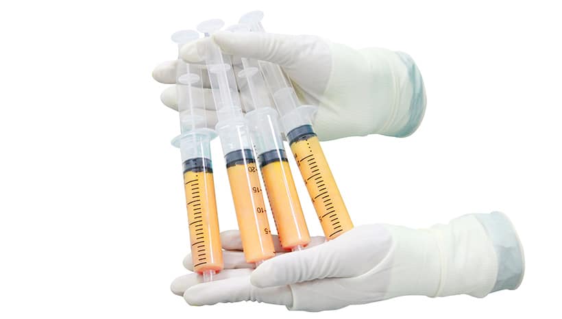
Fat cells that don’t survive after a BBL are resorbed by the body. Dead fat causes local inflammation, with immune cells sweeping up the wreckage over days to weeks. A bit of liquid seeps out through tiny vessels and is reabsorbed. Survivors lodge and provide permanent bulk. This impacts your final shape and can cause bumpy areas or in some instances, the requirement of touch-ups. The bulk of the article touches on risk, timeline, and care to optimize results.
The Clearance Process
Following a BBL, some of the fat cells that are transferred don’t develop their own blood supply and perish. The body identifies these non-viable cells and initiates a complex clearing process that can take days to months. It breaks down the process of your body locating and eliminating dead fat and explains why such removal is important for final form and security.
1. Ischemia
Decreased blood circulation, or ischemia, is the primary culprit in post-graft adipocyte death. When fat is relocated to the buttocks, cells depend on adjacent capillaries to deliver oxygen and nutrients. If new vessels do not stabilize quickly enough, cells go hypoxic and perish. Early indications are pale, cool tissue and swelling or bruising that does not resolve in the expected timeframe. Risk factors are injecting fat in large, dense boluses, overly tight closure, smoking, and patient health problems that impair circulation. Ischemia triggers local chemical cues, including the release of cell debris and apoptotic ‘find-me’ signals that summon immune cells to the graft location and initiate cleanup.
2. Macrophages
Macrophages are big-bodied immune cells that serve as first-line clean-up crews. They smell molecules from dying fat cells and migrate into the region from adjacent blood vessels. Recruitment is chemokine-driven and by the injury-induced increased local blood flow. When they arrive, macrophages bind to and encase pieces of cell membranes and fat droplets. Their work causes mild, acute inflammation. This inflammation aids in debris clearance but can exacerbate swelling and bruising. The right balance of macrophage activity promotes healing. Too much or prolonged inflammation damages surrounding tissue and the cosmetic result.
3. Phagocytosis
Phagocytosis is the consumption and processing of dead cells by macrophages. Steps include recognizing and binding to debris, engulfing it into a vesicle, fusing the vesicle with digestive enzymes, and breaking it down into small components. Quick phagocytosis prevents those oily leftovers from accumulating and creating cysts or calcifications. When phagocytosis is efficient, tissue damage remains minimal and the danger of complications lessens. This clears out potential for infection and drives the area from inflammation to repair.
4. Resorption
After digestion, breakdown products—free fatty acids and glycerol—are transported away via local lymph and blood. Normal resorption occurs over weeks to months and many patients notice that the majority of non-viable fat is cleared by 4 to 6 weeks. The totality of the changes can persist up to 6 months. Approximately 60 to 80 percent of transplanted fat typically sustains. The remainder is resorbed and does not regrow in that location. Utilizing a BBL pillow and not sitting on the buttocks for the first 2 to 3 weeks, up to 4 to 6 weeks if advised, alleviates pressure and is crucial in supporting graft survival.
5. Remodeling
Cleared spaces are replaced by newly formed connective tissue and capillaries. Collagen develops, contours morph and firmness evolves with the rearranging soft tissue. Remodeling defines the long-term shape. Patients who adhere to sitting restrictions and follow post-op instructions report better, more stable results.
Clinical Complications
Clinical complications after BBL occur when injected fat cells don’t survive or act aberrantly. Monitoring and early detection matter because signs can span from minor cosmetic concerns to events that are life threatening. Below is a compact list of symptoms that may indicate a complication, followed by a detailed discussion of three common pathologies: oil cysts, fat necrosis, and calcification.
- increasing redness, warmth, or drainage at injection sites
- new, persistent lumps or nodules under the skin
- severe, worsening pain not relieved by prescribed meds
- shortness of breath, chest pain, fainting or sudden dizziness
- asymmetry, contour irregularities, or skin depressions
- low‑grade fever or other signs of infection
- firm, immobile masses that do not shift with touch
Oil Cysts
Oil cysts are fluid-filled sacs of liquefied fat that collect within the tissue following fat necrosis. They occur when sizable fat grafts lose their blood flow, burst, and leak oil. The oil gets walled off by the body, forming a palpable sac. Clinically, they are felt to be soft or fluctuant initially, occasionally mobile, and may harden if inflamed or infected. On ultrasound, they present as anechoic or hypoechoic cavities. Small, painless cysts can often be observed without treatment. Symptomatic or infected oil cysts can be drained with a needle under imaging guidance or surgically removed. Antibiotics are used if infection is suspected. Recurrent cysts or those that cause contour issues may need to be excised.
Fat Necrosis
Fat necrosis is true death of fat tissue that leaves behind hard, frequently irregularly shaped lumps in the grafted site. Risk factors are fat overfilling a local zone, poor technique, and compromised blood supply. Injecting too much fat into a single plane increases the risk. Post BBL may manifest as tender or non-tender firm nodules, localized induration or hard areas lasting for months. On occasion, skin covering the area may exhibit discoloration or dimpling. Long term effects are firm scarring, persistent lumps, and surface irregularity that sometimes require revision surgery or liposuction to correct. Attention to surgical technique, proper volume distribution, and not jumping into early weight-loss exercise all help mitigate risk.
Calcification
Calcification is the deposition of calcium salts in the sites of previous fat necrosis, resulting in hard nodules. Clinically, calcifications present as small white spots on mammography or CT, and ultrasound may demonstrate shadowing. Most calcifications are benign and only a cosmetic concern. There are some patterns that you want to evaluate to make sure that you’re not dealing with another pathology. Clinically, they can produce palpable hard lumps and dermal roughness which impact contour. Management varies from observation to excision if symptomatic or cosmetically concerning. Avoiding the large areas of necrosis that safe injection techniques prevent reduces its calcification risk.
Procedural Factors
PP). These preferences determine the fate of fat cells that don’t survive after a BBL. The way fat is harvested, cleansed, transplanted, and implanted controls whether cells survive, perish, or are absorbed by the body. The next two sections describe the key steps and their impact, then contrast popular strategies in a rapid-fire bullet list.
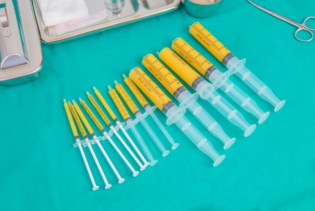
Careful collection counts. Low-trauma liposuction, which involves low suction pressure, small cannulas, and slow, steady passes, minimizes mechanical trauma to adipocytes and the surrounding stromal tissue. When cells are torn or crushed during harvest, they release lipids and inflammatory signals, which boost the likelihood that those cells will die after transfer. Examples of this include power-assisted systems with fine cannulas that can provide better cell integrity than large, high-pressure suction devices.
Its purification before transfer required careful attention. Spinning, filtering, or decanting removes blood, oil, and dead cell debris. If they transfer damaged cells or too much fluid, they increase local inflammation and decrease viable fat survival. A mere gravity decanting or mild centrifugation at low speeds can separate out these contaminants and maintain the cells. For example, a two to three minute low-speed spin often balances cleanup with cell health.
Accurate injection technique and depth placement are paramount. Fat has to be placed in tiny aliquots in several tissue planes so that each droplet is near the surrounding capillaries and can establish a blood supply. Big clumps or shallow puddles have no fast oxygen contact and perish. Injecting in a fan pattern and layering tiny passes maximizes contact with well-perfused tissue and encourages take. Placing micro-aliquots 1 to 2 millimeters apart in deeper subcutaneous tissue improves integration compared with single large boluses.
Control injection volume. Surgeons factor in normal reabsorption, which is frequently 30 to 50 percent, and don’t overfill, which can squeeze local vessels and increase cell mortality. Procedural Factors Staged transfers or modest overcorrection that still honor tissue capacity minimize repeat procedures.
Surgeon experience and donor site play a role. Experience informs cannula selection, manipulation, and positioning. Fat from the lower back or flanks has demonstrated better graft take than fat from fibrous or inflamed areas. Patient factors—smoking, diabetes, poor circulation, or low BMI—reduce viability by compromising revascularization.
Post-operative care is crucial. Early pressure, sitting, or heavy exercise can crush delicate grafts and diminish circulation. Adhering to directions on offloading the region, sleeping positions, and activity restrictions for a few weeks allows cells to regenerate with new vessels.
Compare outcomes by procedural approach:
- Low-trauma harvest and gentle purification with micro-aliquot injection lead to higher survival and a lower complication rate.
- High-suction harvest, minimal purification, and large bolus injection lead to more cell death, greater inflammation, and uneven contour.
- Staged injections with modest volumes lead to predictable and gradual improvement and result in fewer volume losses.
- Single large-session overfilling results in a faster initial shape, but it has a higher risk of reabsorption and fat necrosis.
Recovery Timeline
Post-BBL, your body undergoes an expected healing and fat cell adaptation process. The first few days are about controlling swelling and safeguarding grafted fat. Over weeks and months, the surviving fat cells develop blood supply and integrate. Locally, non-viable fat cells are removed by inflammation and lymphatic drainage, decreasing volume and reshaping during healing.
The majority of fat cell loss and clearance is in the first few weeks. Inflammatory cells and macrophages scavenge the oil, dead cells, and small droplets of fat. This is most active during the initial 2 to 6 weeks, when the body is washing out damaged tissue and fluid. Patients can anticipate most of their initial shrinkage and the majority of early fat loss here. Apparent swelling hits an early peak and then begins to decline. Most of the initial swelling dissipates around 6 to 8 weeks with results becoming crisper as that occurs.
Fat resorption progresses on a general timeline. First week: pronounced swelling, bruising, and tenderness. Soreness can last about 3 to 6 weeks. Weeks 2 to 3: active clearance of dead fat and fluid, gradual reduction in size, continued soreness but less intense. Weeks 3 to 6: patients generally begin to see the shape change as swelling subsides. Avoid prolonged pressure on the buttocks for the first 3 weeks to limit pressure necrosis of transferred fat. Around 4 to 6 weeks: most surgeons allow gradual return to many normal activities. Avoid sitting directly on the buttocks until at least 6 weeks, and avoid sleeping on the back for 4 to 6 weeks to reduce pressure on grafts. Around 6 to 12 weeks: compression garments are typically worn between 3 weeks and 3 months, depending on surgeon guidance. Fat cells that survive start to form stronger blood vessels and stabilize. From 3 to 6 months: continuing settling and improvement. Up to 6 months many patients can see near-final contours. Up to 12 months: complete maturation of the grafted fat and final soft-tissue settling may take up to a year.
Suggested milestones for assessing final results:
- 1 week: immediate post-op swelling and pain baseline.
- 3 weeks: reduction in swelling, beginning of more defined shape, continued garment wear.
- 6 weeks: most swelling reduced, clear for a lot of activities. Careful sitting allowed per surgeon.
- 3 months: substantial settling. Eventually, compare photos to early post-op images.
- 6 months: near-final result for many patients. Review symmetry and volume.
- 12 months: final outcome; graft maturation complete.
During recovery, adhere to your surgeon’s instructions, wear compression garments as directed, and defer heavy exercise until cleared.
Diagnostic Imaging
Diagnostic imaging is central to understanding what happens to fat cells that do not survive after a Brazilian butt lift (BBL). Imaging lets clinicians see soft tissue changes, spot complications early, and tell normal post-operative changes from pathology. Common modalities include X-ray, ultrasound, computed tomography (CT), and magnetic resonance imaging (MRI). Each has specific strengths and limits for evaluating fat survival, necrosis, cysts, infection, or vascular injury.
| Imaging method | Typical use after BBL | Strengths | Limitations |
|---|---|---|---|
| Ultrasound | Assess superficial fat grafts, fluid collections, cysts, guide aspiration | Real‑time, no radiation, bedside use, cost effective | Operator dependent, limited deep tissue detail |
| MRI | Detect fat necrosis, oil cysts, abscess, soft‑tissue edema, vascular compromise | High soft tissue contrast, no ionizing radiation, multiplanar views | Costly, takes longer, potential claustrophobia, contraindicated with some implants |
| CT | Evaluate deep collections, gas (suggest infection), bone or pelvic involvement | Fast, widely available, good for acute surgical planning | Ionizing radiation, limited soft tissue contrast vs MRI |
| X‑ray | Limited direct use for fat; helpful for detecting foreign bodies or displaced implants | Fast, low cost | Poor soft tissue detail |
Ultrasound is frequently the initial imaging for suspected fat necrosis or cysts. It demonstrates well-defined hypoechoic or anechoic lesions when oil cysts develop and heterogeneous, irregular areas when fat necrosis has induced scarring. Clinicians use ultrasound to guide needle aspiration or biopsy of concerning fluid collections. The benefit is bedside use and immediate feedback at follow-up visits.
MRI Offers Comprehensive Map of Grafted Fat and Sequelae. Fat necrosis usually presents as areas of variable signal intensity on T1 and T2 sequences, punctuated by oil cysts with characteristic high T1 signal that drop on fat‑suppressed sequences. Contrast‑enhanced MRI can differentiate inflamed tissue or abscess from simple cysts, thus helping diagnosis and directing treatment. MRI avoids radiation but can cause anxiety or claustrophobia. Open scanners or sedation are options.
CT is useful when deep infection, gas, or urgent surgical planning is suspected. It detects gas in tissue and specifies the spread of collections quickly. The negative is radiation exposure that should be balanced by clinical necessity, particularly in younger individuals.
Imaging guides interventions. Ultrasound or CT can guide percutaneous drain placement or biopsy, and contrast agents improve lesion detection and vascular evaluation. Advances in imaging resolution and faster scans have made many of these procedures more comfortable and accurate. Clinicians must still weigh the benefits against the risks of radiation and patient tolerance.
Surgeon Perspective
Surgeons see the fate of fat cells that don’t survive after a BBL as a combination of evaluation, prevention, and follow-up care. Dead or non-surviving fat is reabsorbed in phases post-surgery. Surgeons evaluate this by looking in the operating rooms, feeling for hardening or masses, and using photos and measurements to monitor volume loss over time. Ultrasound or MRI is employed if there is suspicion of complications such as fat necrosis or fluid collections. If pockets of dead fat develop, a surgeon might suggest conservative care, which includes massage, time, and observation. In symptomatic fat necrosis cases, small procedures may be performed to excise hard nodules.
Surgeons employ a number of tactics to optimize fat survival in a BBL. They harvest fat gently to minimize cell trauma, use low pressure suction, and process grafts to eliminate blood and oil that hamper survival. Injection technique matters: layering small aliquots in many planes increases surface area contact with surrounding tissue, which helps reestablish blood flow. Surgeons map donor and recipient sites meticulously to prevent overfilling, as overfilled grafted fat cannot get to oxygen and perishes. They caution against compression to the new fat for at least a few weeks and often suggest that you avoid any direct pressure and excessive activity for 2 to 4 weeks in order to minimize compression and promote revascularization.
Follow-up is at the heart of a surgeon’s practice. Regular post-op visits in the initial days, two weeks, and at several months allow the surgeon to identify early complications and track fat survival. Surgeons sometimes tell patients that the final aesthetic outcome is not immediate and can take several months to fully develop as the fat establishes its blood supply. Maintaining a daily diary of progress and changes allows both the patient and surgeon to observe patterns in swelling, sensation, and contour. Surgeons stress steady weight control, as significant weight gain or loss can affect graft size and symmetry and sabotage long-term outcomes.
The experience counts. Surgeons with higher case volumes typically have lower complication rates and more predictable outcomes as they fine-tune the harvest, processing, and injection techniques and manage postoperative protocols. Their expertise minimizes overcorrection and limits complications such as fat embolism or necrosis. Junior operators might overlook nuanced indications of bad graft take or recommend subpar aftercare.
Conclusion
Fat that doesn’t survive after a bbl exits the body in obvious manners. The body dismantles dead fat into little fragments. The liver and immune cells clear those. A little bit of fluid and inflammation ensues. It can be hard, sore, or lumpy. Imaging can reveal bumps or fat necrosis. Experienced surgeons employ meticulous technique and follow-up to reduce risk and identify problems early. Recovery habits such as rest, light activity, and no butt pressure facilitate healing and consistent results. For the majority of us, a combination of fat loss and healing results in a shape that’s not quite what you had during week 1. Discuss with your surgeon what to anticipate and the optimal next steps.
Frequently Asked Questions
What happens to fat cells that don’t survive after a BBL?
Fat cells that don’t make it are metabolized by the body. Immune cells come to clean up the wreckage. The leftovers are eliminated via the lymph and urine as metabolic waste.
Can the body reabsorb dead fat cells after a BBL?
Yes. The body reabsorbs and disposes of dead fat cells over time. This can take weeks to months depending on volume and individual healing.
Do dead fat cells cause infection or inflammation?
They can incite some transient inflammation. Good surgical technique and hygiene minimize the risk of infection. These symptoms require immediate medical attention.
How does the clearance process affect final results?
Fat that doesn’t make it after BBL changes shape and volume. Anticipate some volume loss in transferred fat during the initial three months as non-survivors are filtered out.
Will diagnostic imaging show dead fat cells after a BBL?
Imaging may identify fat loss, fluid collections, or complications such as fat necrosis lumps. Ultrasound, MRI, or CT can help distinguish normal healing from complications.
What procedural factors influence fat survival?
Factors that influence fat survival include graft volume, injection technique, blood supply to the recipient site, and surgeon experience. Lower volume and well-dispersed injections increase survival.
When should I contact my surgeon about complications?
Call your surgeon for intense pain, fever, spreading swelling, skin discoloration, or drainage. Early detection avoids severe complications and maintains outcomes.