Key Takeaways
- Extracellular matrix (ECM) scaffolds have been shown to enhance fat graft survival and integration by offering a biomimetic 3D framework that supports cell attachment, reduces resorption, and improves tissue quality.
- Scaffold-derived signals and retained growth factors support adipogenic differentiation and stem cell activity, so pair ECM scaffolds with adipose stem cells to maximize regeneration and clinical outcomes.
- Speedy vascularization is key to graft survival and ECM characteristics like porosity and collagen remodeling induce angiogenesis. This results in improved volume retention compared to traditional fat grafting.
- Select scaffold source and processing thoughtfully, as donor origin, decellularization technique, and material type influence biocompatibility, immune response, and clinical applications.
- Utilize a standardized surgical technique and scaffold handling. Add stem cell enrichment when indicated and track results to minimize fat necrosis and ensure long-term volume stability.
- For clinical application, focus on matching scaffold properties to native adipose ECM, monitor graft survival and complications with objective metrics, and explore ECM scaffolds as platforms for delivering cells or factors in regenerative therapies.
Extracellular matrix scaffold fat transfer clinical use refers to using biological scaffolds to support fat grafting in reconstructive and cosmetic procedures. These scaffolds offer structural support, direct tissue integration, and can enhance graft survival through angiogenesis.
Clinical studies say there is some improvement with volume retention and healing, but they are still developing materials and protocols. The entire body goes over science, approaches, and applications for doctors and users.
Enhancing Fat Grafts
ECM scaffolds can enhance survival and integration of autologous fat grafts, supplying structure, biochemical signals, and a conducive tissue repair microenvironment. They fight typical fat transfer issues like high volume loss and fat necrosis and synergize with ASCs to promote regeneration.
1. Structural Support
ECM biomaterials provide a three-dimensional scaffold that allows adipocytes and progenitor cells to attach, spread, and generate new tissue. This 3D scaffold prevents cells from slumping into dense clusters where oxygen diffusion is limited.
Collagen content and nanofibrous architecture provide mechanical stability. Collagen I and III contribute tensile strength, while elastin and glycosaminoglycans bring elasticity and hydration. Nanofibers simulate native matrix scale, aiding in cell anchoring and load dispersion.
Improved mechanical support prevents early graft deformation and reduces the risk of fat necrosis by maintaining tissue perfusion and space for capillary ingrowth. The main adipose ECM components are collagen types I, III, VI, fibronectin, laminin, hyaluronic acid, and proteoglycans, all of which provide strength and biocompatibility to the scaffold.
2. Cell Signaling
ECM scaffolds serve as reservoirs of biochemical cues that direct adipogenic differentiation and repair. Matrix proteins provide binding sites that alter cell shape and gene expression, directing ASCs toward fat lineage.
Fibronectin and laminin mediate focal adhesion formation and downstream signaling that affects proliferation. Growth factors and cytokines, such as VEGF, FGF, and TGF-β, can be retained within decellularized ECM and released to support regeneration.
These retained signals enhance host response, reduce inflammation, and encourage smoother graft-native interface integration. The outcome is more structured tissue regeneration relative to fat only.
3. Vascularization
Effective, fast vascularization is a must; otherwise, grafts resorb and lose volume. Clinical data demonstrate a 40 to 60 percent loss at six months, with some cases showing nearly a 45 percent mass loss a year.
ECM scaffolds encourage angiogenesis by both supplying porous conduits for endothelial cell penetration and exhibiting matrix-bound angiogenic factors. Scaffold porosity, pore interconnectivity, and collagen remodeling accelerate vessel formation.
Investigations comparing scaffold-assisted to standard grafts describe a faster capillary density increase and superior perfusion, providing early nutrient delivery and limiting ischemic resorption.
4. Stem Cell Niche
ECM scaffolds reconstruct a niche that maintains ASC viability and functionality. The matrix replicates native signaling required for stem cell preservation and regulated differentiation.
A supportive microenvironment enhances ASC retention, proliferation, and adipogenic induction. Histology reveals improved cell populations and engraftment with increasing time when ASCs are combined with ECM, resulting in a more stable tissue structure.
5. Volume Retention
In clinical series, demonstrating that scaffold-enhanced grafts have higher retention and often outperform controls on early and mid-term follow-up. Enhanced retention correlates with decreased resorption and enhanced tissue integration, vascularization, and ASC-driven.
Quantitative comparisons indicate lower percentage losses than traditional fat grafts and mechanically enhanced grafts over the initial six to twelve month period.
Scaffold Origins
Fat transfer ECM scaffolds come from a variety of tissues. Popular tissue sources are human lipoaspirate from liposuction, cadaveric adipose tissue from tissue banks, and porcine tissue from abattoirs. Each source introduces varying biochemical compositions, accessibilities, and processing routes.
Human lipoaspirate is abundant in native adipose ECM proteins and can be manufactured into autologous or allogeneic products. Cadaveric tissue provides larger volumes for standardized allografts. Porcine tissue is the primary source of xenogeneic products due to scale and conserved ECM components.
Donor tissue impacts biocompatibility and downstream clinical application. Fresh autologous lipoaspirate reduces immune risk and is ideal for same session fat grafting. Allogeneic cadaveric products provide off-the-shelf consistency but have increased processing and screening requirements.
Xenogeneic (porcine) matrices must be carefully decellularized to reduce immunogenicity but can still harbor beneficial structural proteins. Selection is based on volume requirements, infection screening, patient immuno-status, and logistics, such as storage and clearance.
Advantages by origin:
- Autologous means minimal immune reaction, immediate use, and a personalized match for ECM cues.
- Allogeneic: Standardized product characteristics, batch testing, off-the-shelf availability.
- Xenogeneic provides an abundant supply, similar mechanical properties, and cost efficiency.
Clinical use weighs these benefits against the risks of disease transmission, immunogenicity, and inconsistency. Regulatory and ethical considerations inform sourcing. Donor screening, consent, traceability, and pathogen testing are mandated for human-derived materials.
Xenogeneic sources need veterinary health records and safety testing. Different regions have different regulations and require sterility, validated decellularization, and bioburden limits. Ethical concerns include donor consent for cadaveric tissue, fair access, and clear disclosure of product source for patient decisions.
Decellularization
Decellularization utilizes detergents (SDS, Triton X-100), enzymes (DNase, RNase), and mechanical agitation to lyse and clear cellular components. Protocols typically mix hypotonic washes, enzymatic digestion, and frequent buffer exchanges to clear nuclear material and minimize ECM destruction.
Sterile rinses and sterilization ensue. So it preserves collagen, laminin, fibronectin, and proteoglycans but strips away the MHC and cell membrane antigens that cause rejection. It retains pore architecture and residual growth factors to direct host cell repopulation.
Decellularization increases host tolerance, decreases inflammation, and promotes adipogenic cell infiltration. Over-processing can debilitate mechanics and strip away necessary matrix cues. Balance is key.
Common decellularization protocols:
- SDS-based detergent cycles with DNase treatment
- Triton X-100 low-concentration washes and enzymatic nucleic acid removal.
- Soak in hypotonic buffer, then use mechanical agitation and nuclease steps.
- Supercritical CO2 extraction combined with mild detergents
- Freeze-thaw cycles prior to chemical processing
Material Types
Biological scaffolds are ECM-derived and bioactive. There are a number of synthetic biomaterials, such as PLGA and PEG, that provide excellent tunable mechanics but do not contain native signaling. Hybrids combine natural ECM with artificial fibers to obtain both signals and strength.
Acellular allografts contribute native proteins and structure. Injectable adipose matrix provides minimally invasive administration. Nanofibrous scaffolds mimic microarchitecture and enhance cell adhesion. Trade-offs are between bioactivity and reproducibility, immunologic risk, and handling characteristics.
Donor-specific ECM differences: porcine and human adipose share collagens I, III, and VI, but quantities vary. Human ECM has more adiponectin-related factors. Material comparison in the table assists clinicians in selecting according to volume, remodeling rate, and regulatory status.
Characterization
- Collagen content: Hydroxyproline assay quantifies types I and III proportions and infers stiffness.
- Lipid residuals: Nile Red staining and GC-MS ensure low lipid for reduced inflammation.
- Mechanical properties: Compression and rheology testing versus native adipose modulus.
- Biologic activity includes growth factor ELISAs and cell seeding assays for viability and adipogenic markers.
Clinical Applications
ECM scaffold-enhanced fat grafting pairs autologous adipose tissue with a bioactive scaffold to enhance graft survival, direct tissue repair, and modulate host response. Here are the major clinical applications, with focused cases and action notes for reconstructive and aesthetic uses, as well as nascent regenerative indications.
Reconstructive Surgery
ECM scaffolds rebuild soft tissue after trauma, tumor, or breast cancer surgery. Tumor or breast volume loss and contour defects persist. In postmastectomy settings, scaffold-augmented fat grafts fill lumpectomy cavities, better contour the chest wall, and lend more reliable support to skin envelope thinning than fat alone.
Some clinical series have reported fewer events of re-absorption when matrix is used, which reduces the number of repeat procedures needed following tumor excision. Scaffold-assisted grafting supports free flap adipose transfers. When small-volume fat is needed to refine flap contour, blending ECM with microfat enhances integration and vascular ingrowth, reducing fat necrosis rates.
For implant-based reconstruction, ECM can cushion thin mastectomy flaps and provide a bed that absorbs supplemental fat with less graft loss. Beyond soft tissue, engineered adipose constructs seeded on ECM exhibit potential for bone and muscle repair. In craniofacial defects, these adipose-ECM composites left next to bone grafts fill dead space and secrete osteogenic factors.
Using similar volumetric muscle loss models, these scaffold-supported fat grafts serve as a pro-regenerative niche that stimulates myofiber growth and restores partial function. Long term benefits are more resilient volume retention and healthier tissue. ECM decreases inflammation, facilitates neovascular networks and directs host cells to remodel the graft into organized tissue instead of cystic fat deposits.
This results in improved contour stability and soft tissue functionality in the long term.
Aesthetic Enhancement
ECM-enhanced fat grafts are used for breast augmentation, contouring, and sculpting in aesthetic and reconstructive procedures where natural feel and integration are the highest priorities. The scaffold enables the volume to hold with fewer touch-ups than with plain fat transfer in some cohorts.
Facial applications exhibit smoother skin texture and less irregularity, specifically in thin soft tissue regions. Versus conventional fat injection, scaffold-assisted procedures generally demonstrate increased graft retention and superior tissue quality on imaging and biopsy. Patients are generally quite satisfied and respond well to the natural feel and longer-lasting results.
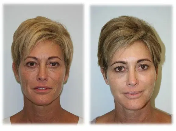
Results are technique-dependent and will vary by donor site and among patients. Common aesthetic indications for ECM scaffold use include:
- Breast contour refinement and small-volume augmentation fills in contour defects after surgery or for slight volume enhancement.
- Taking care of the lost facial volumes — cheeks, nasolabial folds, and tear troughs with less risk of irregularities.
- Hand rejuvenation replenishes dorsal volume and camouflages tendons and veins.
- Scar smoothing and depression correction enhances pliability and weaves into scar beds.
- Buttock and hip contouring provides projection with lower resorption risk.
Surgical Technique
The surgical technique for ECM scaffold-enhanced fat transfer starts with patient selection and site marking and flows into harvest, scaffold preparation, graft enrichment, implantation, and post-op follow-up. Candidates include adult females over 18 years undergoing pre-pectoral breast implant revision or correction of congenital defects, either unilaterally or bilaterally. A recent breast infection within 12 months excludes patients. Follow-up for two years to track safety and outcomes is planned.
Adipose harvest uses tumescent infiltration with an epinephrine solution at a ratio of 1 to 500,000 in normal saline to limit bleeding and ease aspiration. Fat is softly aspirated with a 2.5 mm blunt-tip cannula connected to a Luer-lock syringe, minimizing shear and protecting cell viability. Collected lipoaspirate is washed and processed by low-speed centrifugation or decantation to separate oil and blood while leaving the stromal vascular fraction intact.
Proper graft preparation involves minimal manipulation, no aggressive centrifuge speeds, and keeping it sterile. Scaffold manipulation and seeding ensue. ECM scaffolds come sterile and either dry or hydrated. If dry, hydrate in sterile saline according to the manufacturer’s instructions. Optimize scaffold size and porosity to match the defect so cells spread evenly.
For scaffold seeding, combine processed fat with the scaffold in a sterile field, so fat intersperses the ECM pores. Stem cell enrichment can be incorporated by isolating the stromal vascular fraction or with automated point-of-care devices and then mixing that concentrate with the fat-scaffold composite. Enrichment enhances graft take and vascular ingrowth. Keep track of technique and cell numbers if possible.
Surgical technique is key. Insist on small-volume, multi-plane injections delivered via a blunt cannula that places fat-scaffold composite in thin ribbons and small parcels rather than large boluses that will prevent perfusion. Subcutaneous abdominal placement for investigational protocols: inject AAT and subsequently excise implants during panniculectomy or abdominoplasty at 1 to 18 weeks if needed for study analysis.
In scaffold-guided breast surgery, position the composite pre-pectorally, shape to contour and anchor as required. Plan for convalescence as long as 1 week and advise them regarding restrictions and wound care. Develop and implement a procedural checklist: patient screening (infection history), tumescent solution concentration, cannula size, processing steps, scaffold hydration, cell enrichment protocol, injection volumes per pass, follow-up schedule.
The scaffold is bioresorbable and will be absorbed in possibly as long as three years. These are still ongoing with safety and efficacy scaffold-guided breast studies, monitoring complications and volumetric outcomes up to two years.
Clinical Outcomes
Clinical studies of ECM scaffold-assisted fat grafting describe both improved graft survival and signs of tissue regeneration compared with lipoinjection alone. Early human and animal work demonstrates the scaffold material remains in defects for a minimum of 7 days following treatment and can be detected on histology up to 18 weeks.
Histopathological analysis over 1 to 18 week timepoints reports scaffold persistence, neovascular ingrowth and progressive adipogenesis in several specimens. Volume retention in 6-week-old athymic mice was higher for AAT constructs than standard fat grafts, paralleling increases in viable adipocytes in scaffold groups.
Survival Rates
Clinical and preclinical data demonstrate improved graft survival with scaffold-enhanced fat grafts. In small-animal models, AAT volumes maintained over time outperform scaffold-free controls, with significantly more intact adipocytes on histology at matched timepoints.
Human pilot studies that had explant timepoints at 1, 2, 4, 6, and 18 weeks all reported greater viable cell presence within implants than in surrounding tissue during early weeks. This enhanced survival is associated with both early and strong vascularization into the scaffold, enhanced initial cell viability post implantation, and a biocompatible matrix that supports cell adhesion and proliferation.
Reports cite durable outcomes due to diminished shear and mechanical loss during placement and due to the scaffold’s ability to accommodate host-derived capillaries. Suggest instead to condense these across studies into a survival rate list showing absolute percentage, sample size, and followup time for easy cross-study comparison.
Complication Profile
Common complications in fat grafting include fat necrosis, infection, palpable nodules, and graft resorption. Studies comparing ECM scaffold-assisted grafting note lower apparent rates of fat necrosis and reduced early inflammatory nodules, with fewer cases of late volume loss.
Immune profiling shows comparable absolute immune cell counts within implants versus native adipose for subjects excised between 1 to 6 weeks, and early macrophage shifts toward pro-regenerative phenotypes by weeks 1 to 2. PRA assessments at 4 and 12 weeks against baseline blood draws showed no consistent systemic sensitization in reported cohorts.
Safety data emphasize low immunogenicity of AAT and favorable tissue responses, with most adverse events limited and procedure related.
| Complication | Scaffold-assisted | Standard fat grafting |
|---|---|---|
| Fat necrosis | Lower incidence | Higher incidence |
| Infection | Rare | Rare |
| Graft resorption | Reduced | 40–60% loss typical at 6 months |
| Inflammatory nodules | Fewer, transient | More common |
Beyond Volume
Beyond volume means looking past how much material is placed and asking what it does over time inside the body. In fat grafting, that means assessing long-term outcomes: whether the graft keeps its shape, how it bonds with host tissue, and what biologic changes take place weeks to months after injection. Retention is not just a matter of injected milliliters.
Studies show graft fate depends on host-graft interactions, immune response, and the local tissue environment. Histopathology often documents cellular migration and integration beginning within the first week and continuing through one to eighteen weeks, with clear shifts in cell populations and matrix deposition during that window.
ECM scaffolds transform the local niche. Once adipose tissue is integrated with an extracellular matrix scaffold, the scaffold can enhance tissue quality and elasticity by offering structural support for cellular and neo-matrix deposition. ECM encourages collagen remodeling over scar tissue, which creates softer, more pliant tissue.
That scaffolding maintains shape in the interim before revascularization, minimizing early graft loss to ischemia. Scaffolds encourage angiogenesis and soft tissue regrowth via several mechanisms. With their porous structure and biochemical cues that are retained, they recruit host endothelial cells and promote vessel sprouting.
New vessels reduce hypoxia and allow for sustained graft survival. Biochemical signals in ECM modulate macrophage phenotype toward a pro-repair profile, and that immune shift is crucial. Macrophages clear debris, guide remodeling, and support integration. According to animal studies, there was host cell infiltration, stable graft retention, and neovascularization months after implantation, all indicative of clinical potential.
In addition to volume, ECM scaffolds serve as delivery platforms. They may transport stem cells, growth factors, or small-molecule drugs that regenerate. For instance, seeding scaffolds with adipose-derived stem cells can enhance adipogenesis and vascular ingrowth in preclinical models.
Loading scaffolds with VEGF or PDGF can accelerate vessel formation and enhance early survival. Drug-eluting scaffolds provide an opportunity to locally modulate inflammation or fibrosis without systemic exposure. Evaluation must be rigorous and multimodal.
Imaging and histological staining track cell types, matrix changes, and vessel density. Biochemical assays measure collagen subtypes, cytokine profiles, and markers of adipocyte function. Biomechanical testing assesses tissue elasticity and stiffness, which link to clinical feel and function.
For clinicians and researchers, thinking beyond simple volume means designing grafts and scaffolds with attention to biochemical cues, pore structure, immune response, and targeted delivery to achieve durable and functional tissue restoration.
Conclusion
Extracellular matrix scaffolds provide a framework and support for fat transfer. They assist grafts in maintaining form, connecting to host tissue, and accelerating neovascularization. Research demonstrates improved volume retention and reduced cysts with scaffold. Surgeons utilize porcine or human scaffolds or synthetic blends and put them down with delicate layering and gentle handling. Patients experience a more natural feel and longer-lasting results over months with low risks given proper screening and technique. The cost and supply differ by region. For teams seeking longer-lasting grafts and cleaner contour work, scaffolds present a clear instrument. Explore recent clinical trials, compare scaffold types, and hear from peers to choose the right solution for your practice.
Frequently Asked Questions
What is an extracellular matrix (ECM) scaffold for fat transfer?
An ECM scaffold is tissue-derived. It scaffolds injected fat by delivering structural support and biologic signals that promote cell survival, integration, and remodeling into host tissue.
How does ECM scaffold improve fat graft survival?
ECM scaffolds encourage the growth of blood vessels and reduce tissue strain. This enhances oxygen and nutrient support to transplanted fat, augmenting initial survival and volume persistence.
Where do ECM scaffolds come from?
Scaffolds are commonly derived from decellularized animal or human tissues. For example, dermis or small intestinal submucosa. The processing eliminates cells but retains the structural proteins and signaling molecules.
Are ECM scaffolds safe for clinical use?
If properly processed and approved by regulators, ECM scaffolds have shown good safety profiles. Risks encompass infection and inflammation; however, these aren’t common with the usual surgical care.
Which clinical procedures use ECM scaffolds with fat grafting?
Popular applications are for facial rejuvenation, breast reconstruction and contouring, and soft-tissue defect correction. Scaffolds are utilized when volume retention or tissue quality is enhanced.
What is the typical surgical technique for combining ECM and fat grafts?
Surgeons blend or layer purified fat with scaffold material and deposit it in tiny aliquots. Gentle handling, small-volume placement and careful layering maximize integration and minimize complications.
What clinical outcomes can patients expect after ECM-assisted fat transfer?
In many cases, patients experience increased volume retention, improved tissue texture, and contour stability. Results differ based on patient wellness, defect size, and method, so grounded expectations are necessary.