Key Takeaways
- A pinch test is your fast and non-invasive first line of defense in estimating subcutaneous fat and liposuction candidacy. It must be done consistently to be helpful.
- Only the subcutaneous layer immediately beneath the skin is extractable via liposuction, so a substantial, measurable pinch fold in millimeters or centimeters supports candidacy.
- A hard or firm abdomen to palpation suggests predominately visceral fat, which liposuction cannot address. Use the pinch test to aid in fat type differentiation.
- Standardize body position, tissue grip, measurement tools, and documentation to improve repeatability and accuracy. Repeat measurements at multiple sites for a full assessment.
- Pinch test, snap-back or not, of skin. Bad elasticity puts you in danger of sagging after the procedure and should inform your treatment planning.
- Reserve the pinch test for liposuction candidacy as a companion to objective methods like ultrasound or caliper measurements and practitioner skill and protocol research.
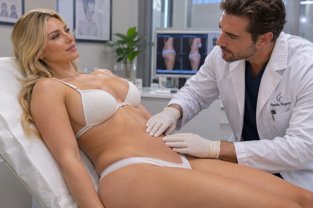
The pinch test for liposuction candidacy is an easy skin-fold check to determine fat and skin elasticity. The pinch test for liposuction candidacy indicates how much tissue you can pinch between your fingers and predicts results from liposuction compared to the necessity of skin tightening. Surgeons use the test in conjunction with your medical history and diagnostic imaging to inform treatment selection and manage expectations. The post below details how the test is conducted, results interpretation, and patient next steps.
Evaluation Basics
The pinch test is a basic, hands-on way to get a rough idea of the thickness of your subcutaneous fat and whether you’re a liposuction candidate. It’s about pinching a fold of skin and subcutaneous tissue between fingers to detect and quantify the extent of soft tissue that can be elevated away from the underlying musculature. The test helps differentiate fat that sits right under the skin and can be liposuctioned from fat that is deeper or bound to organs and cannot be removed safely. When done consistently, the pinch test provides an immediate, non-invasive starting point to evaluation, informing additional evaluation like body composition testing, imaging, or clinical measurement.
Subcutaneous Layer
Subcutaneous is the main fat layer liposuction is aimed at. It lies just below the dermis and above the muscle fascia, creating a soft padding that can frequently be grasped and moved around by hand. This layer needs to be of sufficient thickness to allow for fat removal because if the layer is too thin, suctioning can leave irregularities or contour defects. The pinch test attempts to measure just this subcutaneous compartment and not deeper deposits. In practice, a clinician will quantify this thickness in millimetres or centimetres. For instance, numerous surgeons seek a minimum of approximately 1 to 2 cm of pinchable subcutaneous fat in the area of concern, although acceptable values differ based on technique and target location. The test provides physical tissue feel. Soft, pliable tissue tends to react well to liposuction while fibrous or scarred tissue may be more resistant.
Visceral Fat
- Mostly visceral fat patients are terrible candidates for liposuction because visceral fat sits inside the abdominal cavity around organs and therefore cannot be simply suctioned from the surface. To get it off, you need to lose weight or control it medically, not surgically contour it.
- A hard, unpincheable belly on palpation or a thin, unpincheable layer over a firm belly is often more visceral fat, even if your weight or BMI is high.
- Liposuction in patients whose excess is primarily visceral will not result in significant health or aesthetic enhancement and can lead to unrealistic anticipations. Subcutaneous reduction by itself cannot shrink your waist if the visceral component rules.
- Use the pinch test as one tool to differentiate. If the pinchable layer is minimal but bulk persists, prioritize imaging such as ultrasound or CT, or metabolic evaluation. Counsel patients toward diet, exercise, or medical therapy rather than surgical fat removal.
Assessment Steps
The pinch test evaluates subcutaneous fat thickness to help determine liposuction candidacy. The following subsections break down the practical steps, standardization needs, documentation practices, and a simple checklist to guide clinicians through a repeatable assessment.
1. Body Positioning
Stand at ease, arms hanging down at the sides for the examination. Fixed posture prevents soft-tissue shifts that alter measurements.
Don’t lean forward or back, keep your spine neutral. Insignificant adjustments in the tilt or arm position change the fold geometry and thus can generate different readings. For instance, a patient who has hunched his or her shoulders may exhibit a thinner abdominal fold than when he or she stands.
Request the patient to respirate normally and not to flex. Compare standing to supine positions only when a protocol indicates both and record which was used.
2. Tissue Grip
Pinch a fold of skin and subcutaneous fat between the thumb and forefinger. Skin and fat only, do not pull underlying muscle into the fold.
Grip pressure should be light and consistent. Pinching too tightly squeezes fat tissue and provides artificially low thickness scores. Practice the grip on a few volunteers prior to clinical use to develop a firm hand.
They trained assistants to apply the same grip every time. Marking hand placement on the clinician’s glove or using a tactile cue helps maintain uniform technique across sessions.
3. Thickness Measurement
Measure the fold you pinched with a caliper or ruler and record it in millimeters. Calipers are more precise, particularly for thin folds that are less than 10 mm.
Sample several readings over designated treatment areas, with a minimum of three per location, and average them. Compare values against predefined criteria for safe liposuction candidacy, such as preferred minimum pinch thicknesses of many surgeons in the 10 to 20 millimeter range, depending on location and technique.
Note device type, calibration status, and measurement units in the chart to enable subsequent comparison.
4. Muscle Tension
Request patients to relax abdominal and regional muscles at the time of measurement. Tension minimizes perceived fat fold thickness and can conceal actual subcutaneous volume.
Palpate first to feel the spasm. If there is tension, stop and let the patient breathe and relax. Then take the measurement again.
Record muscle tone notes. Either chronic hypertonicity or recent exercise can influence repeat measures.
5. Results Review
Cross-reference measured thicknesses with minimum safety thresholds listed in your protocol. Indicate findings in a quick report by site, mean pinch value, device, and patient position.
Identify target areas with adequate subcutaneous fat and flag zones that are too thin for safe liposuction. Use the data to plan further imaging or to inform the patient discussion about expectations and risks.
Create and store a standardized checklist for every assessment to ensure consistency and enable comparisons over time.
Skin Elasticity
One of the telltale signs of youth is skin’s ability to bounce back after having been pinched. Skin elasticity is important for liposuction as well because loose or inelastic skin might not adapt to the new shape post fat extraction, resulting in sagging or requiring further treatments. Testing elasticity provides a functional perspective on probable surgical results and can help manage expectations.
- Lack of elasticity can cause sagging, uneven contours, extra folds, obvious wrinkling and slow wound healing, resulting in skin excision, longer recovery and less predictable aesthetic outcome.
Pinch and release the skin you’re measuring and observe its snap-back speed and quality. Apply a firm but gentle pinch with your index finger and thumb, pull the skin fold away from the underlying tissue, hold a second, then release. Note two aspects: how quickly the skin returns, which is the snap-back time, and whether it returns smoothly or shows wrinkling, dimpling, or delayed recoil. Compare multiple sites: abdomen, flanks, medial thighs, lateral thighs, and upper arms. For instance, abdominal skin that takes longer than two to three seconds to snap back into place after pinching and exhibits surface creases indicates low elasticity and greater susceptibility to sag following liposuction. Thigh skin that snaps back in under a second demonstrates good elasticity and a post-op contour that will likely be smoother.
Acknowledge that poor elasticity can create sagging after liposuction. Patients with chronic weight fluctuations, severe sun damage, those of advanced age, smokers, or those specifically genetically predisposed will demonstrate worse recoil. Previous pregnancies will have thinned and stretched the abdominal skin so it will almost certainly not retract completely. In these cases, combine the pinch test with clinical judgment about skin quality. Look for stretch marks, loss of dermal thickness, and soft tissue descent. Discuss options such as staged liposuction with skin-tightening procedures, energy-assisted liposuction that may provide modest tightening, or planned excisional procedures like abdominoplasty when laxity is marked.
Record elasticity findings with fat thickness. Mark the site, pinch-test duration in seconds, qualitative observations such as smooth recoil, wrinkled, tethered, and fat thickness in millimeters either with calipers or ultrasound. Example entry: “Right flank: pinch recoil 1.2 seconds, smooth; subcutaneous fat 18 millimeters by ultrasound.” Regular documentation helps with surgical planning, informed consent, and post-op comparisons. When available, utilize photographs with standardized positioning and scale markers.
Mix pinch-test results with other evaluations, such as skin quality, patient age, BMI, and lifestyle, to create a clear strategy and advise patients on realistic results.
Critical Caveats
The pinch test provides a rapid approximation of the thickness of subcutaneous fat by quantifying the amount that can be pinched between your fingers. It provides helpful surface knowledge but suffers from significant shortcomings. Here are the key caveats doctors and patients should be aware of prior to incorporating the pinch test into a liposuction candidacy evaluation.
Acknowledge that the pinch test does not measure internal (visceral) fat
The pinch test only samples subcutaneous fat which lies right under the skin. It can’t detect visceral fat, which lurks around internal organs inside the abdominal cavity. One can have a modest skinfold measurement yet be harboring dangerous levels of visceral fat, which influences health risk factors and can change one’s aesthetic objectives. For instance, two patients with the same pinch at the waist can display wildly different waist circumference and metabolic profiles if one has elevated visceral fat. Dependence on the pinch test alone can thus give both surgeon and patient false impressions about overall body composition and achievable liposuction results.
Warn that improper technique can lead to inaccurate assessments
There are important caveats. Pinch location, finger placement, patient posture, and force applied all change outcomes. Pinching over a muscle bulge or scar or when the patient is tense can provide falsely low or high readings. Use consistent landmarks, such as a certain distance away from the umbilicus for abdominal measurements, and exert light, uniform pressure to prevent tissue from being compressed. Training and calibration among examiners alleviate this variability. For instance, a surgeon ought to perform the pinch, cause the patient to relax, and then perform it two or three more times, averaging results to get a more dependable number.
Note that individual anatomy and skin conditions may affect results
Skin laxity, thickness, stretch marks, previous surgery and edema affect the way fat pinches. Older adults can sometimes have thinner, less elastic skin, so small fat deposits may feel larger. Scar tissue from previous liposuction or abdominal surgery can adhere tissue planes, rendering pinches irregular. Lipedema is characterized by hard, painful fat that feels different on touch than normal subcutaneous fat. Assume that anatomy and skin condition will bias pinch values and read them in concert with visual inspection and patient history.
Stress that the pinch test is only one part of a comprehensive evaluation
A full candidacy assessment includes medical history, body-mass distribution, imaging when needed (ultrasound or MRI for uncertain cases), skin quality evaluation, and discussion of goals and risks. Use the pinch as a quick bedside metric, not a decision rule. Combine it with objective measures like circumference, weight trends and, when appropriate, metabolic screening.
Clinical Alternatives
Clinical alternatives to the pinch test for determining liposuction candidacy. Here’s some background, followed by two popular alternatives: ultrasound scans and caliper metrics that provide alternative benefits. We start with where alternatives might be preferred, followed by a detailed coverage of each method with side-by-side comparisons, examples, and tips for combining multiple approaches.
- When patients have thick or lax skin and it is not clear how deep the fat is.
- When prior surgery or scarring alters tissue feel
- When visceral fat or organomegaly is a concern
- When accurate volume estimates are necessary for planning or billing.
- For example, monitoring minor fluctuations post weight loss or therapy.
- In medicolegal or research settings that demand objective documentation
Compare accuracy and objectivity: ultrasound is more objective than the pinch test and gives direct imaging. Calipers are more reproducible than manual pinch alone, but are user-dependent. The pinch test is rapid and practical in most clinics. Clinical alternatives add precision where required. Best results are achieved by combining the methods—use pinch for screening, ultrasound for localization and volume, and calipers for longitudinal tracking.
Ultrasound Scans
| Feature | Subcutaneous Fat | Visceral Fat |
|---|---|---|
| Visibility on ultrasound | Clear hypoechoic layer under skin | Often not directly visible on standard abdominal wall scans |
| Measurable thickness (example) | 0–50+ mm depending on site | Indirect assessment via organ displacement, measured via specialized protocols |
| Clinical utility | Direct measurement for local planning | Assesses intra-abdominal risk but needs different probes/protocols |
Ultrasound can vividly display subcutaneous fat thickness and the fat-fascia plane. That distinction matters: liposuction removes subcutaneous fat, not visceral fat. Millimeters of thickness at specific sites are quantitative measures that help set realistic volume targets. In challenging or borderline cases, such as post-bariatric skin laxity or previous surgery with scar tissue, ultrasound eliminates the guesswork. For example, a patient with 8 mm subcutaneous fat at the flank likely needs limited suction. A patient with 40 mm requires broader planning and possible staging.
Caliper Metrics
| Site | Typical adult male norm (mm) | Typical adult female norm (mm) |
|---|---|---|
| Triceps | 10–15 | 15–25 |
| Subscapular | 12–18 | 14–22 |
| Suprailiac | 10–20 | 18–30 |
Go with standard sites—triceps, subscapular, suprailiac, abdomen—for uniform measurements. Caliper measurements are matched against population standards to categorize low, normal, or high subcutaneous thickness. Record values at baseline and at follow-up to track changes after diet, exercise, or procedures. Calipers are inexpensive and portable, but they require training to minimize inter-operator variability. Repeated measures by the same clinician improve reliability. Considering caliper trends in conjunction with imaging and clinical exam generates the most secure plan.
Practitioner Perspective
The pinch test is a quick bedside method to get a guesstimate of subcutaneous fat thickness and assist in determining liposuction candidacy. It involves pinching the skin and underlying fat between fingers or calipers to measure fold thickness in millimeters. The test can reveal if fat is primarily superficial and pinchable, which is the type that responds well to suction-assisted procedures, or if tissue is fibrous, adherent, or visceral-dominant, which all make liposuction a poor fit.
Dependability and restrictions of the pinch test. The pinch test provides good directional guidance, but it has obvious boundaries. Inter-operator variability is high. Different clinicians will record different values on the same patient unless they use a caliper and strict technique. It’s sensitive based on where on the body it’s taken. For instance, a 20 mm pinch at the supraumbilical doesn’t equate to 20 mm at the flank because tissue composition is different. The test cannot sense deep or visceral fat and cannot measure the skin’s elasticity, which plays a big role in contour results after fat removal. Scar tissue, prior surgery, and major edema can distort readings. Examples: a patient with a 25 mm flank pinch but poor skin recoil may have visible irregularity after liposuction. A 15 mm upper arm pinch in an athletic person might reflect thin subcutaneous fat with more fibrous tissue, making suction less effective.

Practitioner’s point of view. Experience informs how we read the pinch test data. Experienced practitioners cultivate a tactile intuition for tissue texture, such as soft, malleable fat versus hard, fibrotic tissue, and combine pinch numbers with visual examination and patient history. They know what body zones require repeated measures and how to standardize hand placement. For example, a seasoned clinician will verify standing versus supine, compare bilateral sites, and correlate with BMI and fat distribution. Inexperienced clinicians might lean too heavily on the raw numbers and overlook contextual hints like prior weight loss or lax skin.
Combining clinical judgment with objective tools best practice pairs the pinch test with objective measures. Calipers are used for consistent folds, ultrasound is used for depth and fascial planes, and standardized photography is used for documentation. Ultrasound can expose deep fat pockets or septations that the pinch test misses. Mixing techniques helps clear up confusion. For example, a moderate pinch accompanied by ultrasound confirming superficial fat encourages you to proceed, while a similar pinch but ultrasound confirming thick deep layers has you reconsider.
Continued practice to get better at the pinch test. Skill improves with structured training, including workshops using standardized patients, supervised clinics, and regular audits of pre-op pinch readings against intraoperative findings and outcomes. Simulators and video review help instill consistency. Regular calibration sessions, in which teams compare caliper readings, minimize inconsistency and increase the general standard of evaluation.
Conclusion
Pre-lipo pinch test provides a definitive, rapid screen of skin and fat. It indicates a person’s loose fat amount and implies how the skin will lay post-procedure. Apply the test alongside a comprehensive exam, photos, and patient objectives. Note the limits: age, sun damage, big weight shifts, and medical issues change results. For mild cases, try non-surgical skin tightening or fat freezing options. Collaborate with a skilled practitioner who documents results and establishes achievable objectives.
If you want a handy checklist, step-by-step guide, or sample consult notes, just ask and I’ll send you one you can use immediately.
Frequently Asked Questions
What is the pinch test for liposuction candidacy?
The pinch test estimates how much skin and fat you can pinch up between fingers. It lets us know if fat removal alone will do the trick or if skin will require additional tightening procedures.
How do I perform the pinch test at home?
Pinch the area in question between your thumb and fingers, lifting skin and fat away from underlying tissue. Mark thickness in millimeters or as small, moderate, or large. Duplicate in multiple locations for contrast.
What does a thin pinch result mean?
A thin pinch indicates little subcutaneous fat. Liposuction might not offer visible contouring and can result in dimpled skin or excess skin.
What does a thick pinch result mean?
A fat pinch with high thickness means you have enough fat for liposuction. Outcomes tend to be smoother if the skin is sufficiently elastic.
How does skin elasticity affect the pinch test result?
Good skin elasticity means the skin bounces back once the fat is taken away. Bad elasticity increases the risk of sag and might necessitate skin tightening or other treatments.
Are there limitations to the pinch test?
Yes. The pinch test is a rough screening check. It can’t evaluate deeper fat, muscle tone, or overall body proportions. You need a professional’s opinion.
When should I see a practitioner after doing a pinch test?
If your pinch indicates redundant fat or lax skin, schedule a consultation. A good surgeon will blend the pinch test with medical history, exam, and imaging to create a safe plan.