Key Takeaways
- Ultrasound cavitation utilizes targeted high-frequency acoustic waves to generate microscopic bubbles that physically break down fat cell membranes, emulsifying localized fat for metabolic elimination via the body’s lymphatic and hepatic networks. Follow suggested post-treatment hydration and movement to assist clearance.
- Frequency and intensity dictate both depth and effectiveness, with lower frequencies targeting deep fat and higher frequencies targeting more superficial layers. Practitioners adapt settings to adipose depth and do not use excessive intensity for overheating.
- Tissue characteristics like density, hydration, and collagen influence cavitation efficacy. Pre-treatment evaluation with skinfold measurements or imaging informs dosimetry customization for safer, more efficient results.
- Clinical and laboratory data demonstrate detectable reductions in subcutaneous fat layer thickness, triglyceride release from fat cells, and enhanced skin texture and appearance after several treatments. Results take time and differ from person to person.
- Observe acute metabolic symptoms such as a temporary triglyceride spike, localized swelling, and mild erythema. Follow longer-term markers like subcutaneous fat mass, hepatic health, and overall body composition to validate safe systemic clearance.
Ultrasound cavitation and fat cell disruption science examines how highly concentrated sound waves rupture fat cell membranes. It covers the mechanism, typical energies in the kHz and MHz ranges, and measurable effects such as adipocyte volume reduction and local circumference change.
The study includes device types, number of treatments, and immediate and longer-term fat loss. Safety data and common side effects are reported.
The body of the article covers studies, protocols, and patient selection.
The Cavitation Mechanism
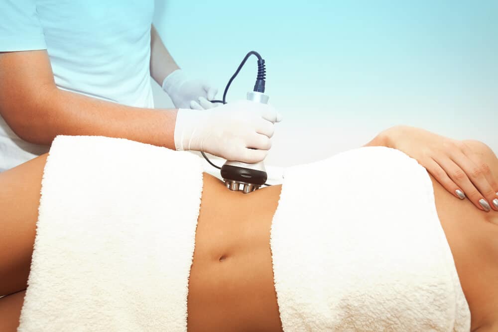
Ultrasound cavitation is the process by which ultrasonic waves create pressure changes in fat cells that result in their destruction. In non-invasive body contouring, focused ultrasound energy targets subcutaneous fat layers while sparing the skin and surrounding tissues.
About The Cavitation Mechanism
This section dissects how devices generate waves, create bubbles, rupture cells, release lipids, and facilitate systemic clearance.
1. Wave Generation
Ultrasound probes pass over the skin of selected subcutaneous fat deposits. Experts pick MHz ranges, with lower frequencies going deeper and higher frequencies acting more superficially. Frequency and intensity dictate both depth and amount of mechanical stress on fat, so machines designed for cavitation employ tuned ranges to maximize adipocyte targeting.
Proper delivery matters; misapplied energy can heat or injure nonfat tissue. Typical sessions last 45 to 75 minutes, enabling precise exposure throughout the treatment area.
2. Bubble Formation
Acoustic cavitation produces micro-bubbles in the adipose stroma. These microbubbles rapidly expand and implode, creating localized pressure peaks that disrupt adipocyte membranes.
Bubble action is more robust in well-hydrated tissue and regions with greater fat content, accounting for differences between patients and body locations. Multiple ultrasound sessions amplify cumulative bubble impacts, and several treatments are generally planned to achieve clinical circumference decreases of approximately 2 to 4 centimeters.
3. Cell Disruption
Collapsing cavitation bubbles micro-rupture fat cell membranes, resulting in selective adipocyte lysis. That mechanical action pushes triglycerides and fatty acids out of cells.
Ultrastructural studies reveal disrupted lipid vacuoles and cytoplasmic changes consistent with membrane poration and rupture after treatment. Nearby adipocytes and collagen fibers remain generally unscathed, demonstrating the relative tissue selectivity of ultrasonic cavitation compared to thermal modalities such as RF.
4. Fat Release
Liquefied fat and triglyceride droplets are extravasated into the adipose interstitium. Stray lipids, mostly triglycerides, free fatty acids, and glycerol, are liberated into the local interstitium and then into lymphatic vessels.
This discharge is evidenced by triglyceride assays and by subcutaneous biopsies following treatment. About: The Cavitation Mechanism Cryolipolysis and ultrasound cavitation both target selective fat cell death. Cavitation achieves this through the mechanical collapse of bubbles instead of cold-induced apoptosis.
5. Systemic Clearance
Your body burns and excretes liquefied fat via lymphatic delivery, liver absorption and oxidation. Thirst and normal lymphatic function, along with good hydration and regular lymphatic function, help it clear rapidly.
If you’re dehydrated, it’s slow. Several treatment studies note alterations in liver lipid handling and small improvements in some serum lipid markers, although temporary elevations in blood triglycerides can happen. The full visible results usually manifest six to twelve weeks following the last session, and mild, temporary side effects such as numbness or redness are common.
Influencing Factors
Ultrasound cavitation results are contingent upon the machine, tissue, and patient factors that collectively contribute to cavitation generation, energy delivery, and fat elimination. The subsequent subtopics dissect how frequency, intensity, and tissue properties influence efficacy and safety and how patient habits and strategies impact outcomes.
- Ultrasound frequency: low (20 to 40 kHz) versus mid (40 to 100 kHz) versus high (greater than 1 MHz) ranges
- Ultrasound intensity is reported in watts per square centimeter. Higher values increase mechanical stress and heat.
- Tissue properties include adipose density, collagen content, hydration status, skin thickness, and perfusion.
Frequency Impact
Lower ultrasound frequencies penetrate deeper into tissue, so they reach thicker subcutaneous pads and peri-visceral fat more easily. For example, 20-40 kHz devices generate bigger cavitation bubbles with higher collapse energy, which can breach larger adipocytes and cause more mechanical stress to adjacent structures.
Higher frequencies in the 1 MHz range have shorter wavelengths and shallower penetration, therefore they are ideal to sculpt superficial fat and treat skin texture through collagen remodeling stimulation. Frequency selection alters bubble size distribution and the formation and collapse rate of bubbles.
Choose lower frequency for deeper liporeduction and higher frequency for surface contouring. Adjust the setting according to the measured fat thickness.
Intensity Levels
Higher intensity increases the pressure amplitude of the acoustic wave, which enhances both cavitation likelihood and the mechanical stresses on adipocyte membranes. That can amplify fat disruption and increases tissue heating and burn risk.
We wanted to identify the intensity window that best optimizes adipocyte disruption while maintaining skin and subcutaneous tissue temperatures below safe thresholds. Intensity can be reduced over delicate spots such as the inner arm and increased for more dense flanks, making spot-specific adjustments.
Professionals should evaluate each session and establish safe margins. Patients should abstain from NSAIDs such as ibuprofen for two weeks prior to treatment to minimize bleeding risk and promote predictable tissue response.
Tissue Properties
Tissue density, hydration, and collagen framework alter the propagation of ultrasound and the formation of cavitation. Well-hydrated tissue more uniformly conducts sound and flushes lipid spillage; therefore, patients are encouraged to consume fluids post-treatment to facilitate metabolite removal.
Dense adipose or loose skin areas need distinct protocols. Dense fat may need higher energy or repeated sessions, while loose skin may benefit from combined frequency and skin-tightening approaches. Basic tools such as skinfold calipers and BMI measurement are perfect candidates.
Individuals typically have a BMI between 18.5 and 24.9 and are within approximately 7 kg of target weight. Unfortunately, smoking compromises microcirculation and healing, which is why non-smokers tend to be better candidates.
Keep in mind that results are contingent on continued healthy eating and reasonable expectations. Cavitation is not a weight loss alternative and works suboptimally at BMIs of 30 or higher.
Scientific Evidence
Ultrasound cavitation technology has research spanning from in vitro studies to clinical trials, with the ex vivo and in vivo work demonstrating consistent fat reduction outcomes. Lab work with skin explants and adipose biopsies reveals changes at the cellular level, and human trials quantify subcutaneous fat volume, circumference, BMI and skin appearance. Your body flushes treated fat over a period of weeks, so the majority of changes are evident 6 to 12 weeks after your last session.
Laboratory Studies
Ex vivo experiments demonstrate adipocyte necrosis, apoptosis, and evident lipid leakage following ultrasound exposure. Treated tissue frequently exhibits membrane rupture and intracellular fat escape on electron microscopy.
Ultrastructural analysis reveals disrupted adipocyte membranes and reduced mean adipocyte size. All three note apparent pitting and loss of cell integrity for samples exposed to the same cavitation frequency and power used clinically.
Triglyceride release and quantifiable fat layer thinning are typical readouts. In some explant models, they measure free fatty acids and glycerol in nearby fluid, confirming lipid release from adipocytes following cavitation pulses.
Laboratory models allow researchers to examine particular ultrasound parameters in a controlled setting. Factors including frequency, intensity, pulse length, and focal depth are calibrated to optimize cell disruption while preserving dermal and vascular tissues. This work informs clinical device settings and safety margins.
Human Trials
Clinical trials document impressive decreases in subcutaneous fat thickness and body fat area following courses of treatment. Measured changes range from circumference drops frequently in the 2 to 4 cm range for treated areas, to one-session fat cell reductions between approximately 14.7 percent to 28.5 percent reported.
A series of treatments over two to eight weeks generally makes skin firmer and smoother, reduces the look of cellulite, and decreases abdominal circumference. Most protocols utilize six to twelve sessions, each session lasting approximately forty-five to seventy-five minutes, spread out over days to weeks to permit slow clearance of liberated lipids.
Ultrasound imaging and other ultrasonography measures confirm these reductions in fat layer thickness. Scientific evidence shows that objective scans before and after the treatment course reveal thinning in subcutaneous layers that corresponds with clinical circumference measures.
Patient reported outcomes prioritize improved shape and visible sculpting. Users typically observe incremental enhancement, with the majority experiencing the greatest transformation six to twelve weeks post-treatment, coinciding with the body’s natural fat processing and elimination timeframe.
Key clinical studies and findings:
- Randomized trials demonstrate a 2 to 4 cm circumference reduction of treated areas.
- Single-session fat loss reports range from 14.7% to 28.5% by cell count.
- Multi-session studies involving six to twelve sessions demonstrate persistent thinning of subcutaneous fat on ultrasound.
- Safety data is consistent with FDA approval and shows low rates of serious adverse events.
- Patient surveys reporting improved contour and cellulite reduction.
Metabolic Aftermath
Ultrasound cavitation initiates an immediate and delayed metabolic response in the adipose cells. Ruptured adipocytes release triglycerides that enter interstitial fluid and drain through lymphatics. Some are locally bound and reused, while others enter circulation and are processed by the liver and kidneys.
The metabolic chain extends from minutes to months and includes fluid shifts, immune clearance, and slow tissue remodeling.
Short-Term Effects
Immediate tissue reactions encompass adipocyte volume loss and a discharge of triglycerides into circulation, which can be detected by biochemical and imaging assays. Local swelling, mild redness and temporary skin flaccidity may ensue a session; these are indicative of fluid shifts and the transient depletion of cellular volume.
Most patients bounce back swiftly since it’s a noninvasive procedure and discomfort is typically minimal. Temporary numbness or bruising may arise, as with other fat-zapping treatments such as cryolipolysis.
Short-term fat reduction is detectable by ultrasound and plicometry and can often be observed in as little as 6 to 12 weeks, although the timing varies from person to person.
Long-Term Effects
Multiple ultrasound cavitation treatments result in a cumulative fat loss and net reduction of adipose tissue mass over a period of weeks to months. Long-term follow-up studies show many patients maintaining weight and displaying decreased markers associated with ectopic fat deposition, potentially reducing fatty liver risk over time when combined with lifestyle modifications.
These changes in shape, skin, and cellulite can be maintained for months after treatment, while macrophages continue to clean up and tissue remodeling occurs. Apoptotic and necrotic remains are incompletely consumed by macrophages over the course of about two to six months in similar treatments, so visual and metabolic shifts may arise gradually.
Lifestyle factors, such as diet, hydration, and activity, very much impact longevity; a healthy, balanced diet and regular exercise promote longevity.
Checklist: Monitoring metabolic markers and changes after ultrasound treatment
- Triglyceride levels: measure baseline and at short intervals to detect transient rises and return to baseline.
- Liver enzymes: track for hepatic processing stress if circulating lipids increase.
- Renal markers and urinalysis: ensure kidneys handle lipid by-products. Sufficient hydration is essential.
- Body composition: Use ultrasound scans and plicometry at six and twelve weeks to quantify fat loss.
- Clinical signs: Record local swelling, redness, numbness, or bruising and any delayed reactions within two weeks.
- Functional measures: note changes in insulin sensitivity or metabolic panels during long-term follow-up.
Practical steps to support metabolism post-treatment include drinking at least 2 L of water daily, performing mild aerobic exercise such as brisk walking within 24 hours, and considering lymphatic drainage massage, either manual or radiofrequency-assisted, to speed free fatty acid clearance.
The Acoustic Window
It’s called the acoustic window, the optimal window through which ultrasound waves can be delivered at a specific frequency to targeted fat layers and strategically disrupt fat cells. For ultrasonic cavitation, such a window typically lies between 40 kHz and 60 kHz, a band that optimizes energy delivery to adipocytes while minimizing it by skin and muscle.
Waveform, transducer design, and tissue type shift the window a bit. Skin thickness, subcutaneous fat depth, and body composition can all alter effective penetration and cavitation thresholds. Ultrasound imaging is frequently used to verify alignment and ensure the beam will pass through a clear path to the fat layer before energy is delivered.
Tissue Selectivity
Focused ultrasound energy at the acoustic window level preferentially targets adipocytes since fat has different acoustic and mechanical properties than dermis and muscle. At 40 to 60 kHz, microbubbles form within the lipid-rich cells, leading to localized mechanical stress and membrane rupture, whereas dermal fibroblasts and epidermal layers absorb less energy and remain intact.
This selectivity reduces collateral damage and facilitates quicker healing, with numerous patients experiencing little to no downtime post-therapy. Good targeting mitigates the risk of harming capillaries and microvasculature that course within and just beneath the fat layer.
When the beam is focused on a subcutaneous pocket, microbubble activity localizes in adipose lobules and spares adjacent microvessels, reducing bruising and vascular damage. That precision permits treatment over standard areas—abdomen, flanks, thighs, and arms—because the system can be calibrated in depth and focal size to correspond with local anatomy and fat thickness.
Safety Margins
Observe safety margins from bone and internal organs. Bone reflects and focalizes ultrasound and causes local heating, while organs can be damaged if energy penetrates too deeply. Professional cavitation units have options for focal depth, pulse length, and total energy distribution so providers can keep exposure safely bounded within the acoustic window.
Many devices have built-in cutoff thresholds and transducer cooling to shield your skin. Keeping an eye on tissue temperature and receiving direct, real-time feedback from patients are both practical approaches to prevent overheating and burns.
Real-time or intermittent ultrasound, prior to and during therapy, ensures that the acoustic window is still in alignment and that energy is not impacting any unintended structures. Staying within recommended safety margins and using imaging verification reduces adverse event rates and can aid in maintaining consistent results.
Beyond The Hype
Ultrasound cavitation applies targeted sound waves to induce microbubbles in fat cells, leading to their membranes’ rupture and lipids’ release into surrounding space for removal. It is confirmed to reduce localized fat and girth, but results are slow and variable.
Realistic Outcomes
Most patients witness modest decreases in subcutaneous fat volume and frequently a more toned skin appearance following a course of treatments. Clinical trials report measurable circumference reductions, typically 2 to 4 centimeters per treated region, according to a 2014 paper in the Journal of Cosmetic and Laser Therapy and a 2020 systematic review that found the method effective and safe for localized fat reduction.
You’re not going to lose dramatic amounts of weight with noninvasive ultrasound, as the procedure attacks subcutaneous deposits, not the deep visceral fat around your organs. It’s no replacement for clinical weight-loss procedures. Patients looking for body sculpting, not massive weight loss, are the best candidates.
Ultrasound cavitation is most effective when used as a complement to lifestyle interventions. A good diet and exercise keep those contour changes longer. A series of treatments, generally six to 12 spaced roughly a week apart, is typically needed. The results tend to show fully over six to 12 weeks, with benefits lasting even beyond the last session.
- Modest circumference and volume loss: expect reductions of 2 to 4 centimeters in treated areas, often spread across sessions.
- Improved skin texture: some tightening or a smoother appearance can follow fat disruption.
- No quick fix for visceral fat. Internal fat and large weight loss are not addressed.
- Multiple sessions and lifestyle upkeep: Treatments work best when paired with diet, hydration, and exercise.
- Low downtime and minimal scarring are advantages over surgery, yet they are less dramatic than invasive methods.
- Variable outcomes: Individual biology and adherence to aftercare change results.
Patient Variability
Age, gender, baseline adiposity, and skin elasticity determine the response to cavitation. Younger skin with great elasticity will bounce back better, while older skin might not appear as tightened even with comparable fat loss.
Some patients experience quicker or more significant decreases, while others creep along. How fast released lipids clear tissue and enter circulation to be processed depends on hydration, metabolic rate, and other factors.
What you do after treatment counts. Stay away from alcohol, excess carbs, and sugars for at least 48 hours to assist clearance and minimize re-depositing. Personalized plans that tailor energy delivery, number of sessions, and aftercare produce optimal results.
Future Directions
Focused ultrasound might become more precise, allowing clinicians to target layers more selectively and minimize collateral impact. Pairing cavitation with other noninvasive modalities, like cryolipolysis for instance, may optimize contouring results by leveraging synergistic mechanisms.
Other studies are testing systemic and metabolic effects of repeat sessions and expanding utilization beyond aesthetic uses, such as focal fatty liver type approaches or for localized adiposity associated with specific medical conditions. Safety and long-term data will inform clinical adoption.
Conclusion
Ultrasound cavitation is a procedure that employs focused sound to create microbubbles in adipose tissue. Those bubbles burst and strain fat cell walls. Research indicates that this ruptures cell membranes and releases fat. Your body goes to work putting that fat into circulation and your liver clears a lot of it out. Results depend on device strength, treatment duration, and physique. Risk remains low but real. Minor bruising, pain, and temporary swelling occur in certain research. Sustained weight loss requires diet and exercise. For a noticeable difference, combine treatments with consistent habits. For instance, include two energetic 30-minute walks a week and monitor protein intake at meals. Discuss objectives and tests with a clinician prior to a treatment series. Find out more and book a consult if you’re interested in next steps.
Frequently Asked Questions
What is ultrasound cavitation and how does it disrupt fat cells?
Ultrasound cavitation works with focused sound waves to form microbubbles in tissue. As the bubbles collapse, they mechanically stress the fat cell membranes and trigger them to release their stored lipids without surgery.
Is the fat removed or just redistributed after treatment?
While the majority of liberated lipids are processed by the body, there can be some short-term redistribution. With the right hydration, exercise, and lymphatic support, the body flushes the lipids rather than storing them in another area.
How effective is cavitation for reducing body fat?
There are clinical studies that demonstrate modest localized fat reduction, on the order of a couple millimeters to a couple centimeters per treated area. Outcomes differ from device to device and depend on the treatment schedule and individual variables such as BMI and lifestyle.
Are ultrasound cavitation treatments safe?
Cavitation, when performed by trained professionals using approved devices, is generally safe. Mild side effects include redness, bruising, or temporary sensitivity. Contraindications are pregnancy, implants, or active infections.
How many treatments are usually needed to see results?
The majority of protocols suggest three to eight sessions spaced roughly one to two weeks apart. You can see results after the second or third session, with more coming over the next few weeks as your body metabolizes the released lipids.
Does cavitation replace diet and exercise for long-term fat loss?
Cavitation addresses those nearby fat pockets. Long term fat control needs healthy eating, exercise, and weight-control habits to keep it off.
What scientific evidence supports cavitation’s metabolic effects?
Peer-reviewed published papers describe elevated free fatty acids in circulation post treatment and localized fat volume reduction. Evidence quality is variable, with larger and longer-term randomized trials still limited but increasing.